Tooth anatomy:
The crown is above the gum line, the root below. The crown is covered with enamel, the hardest substance in the body. Dentin makes up the bulk of the tooth, inside it is hollow with nerves and blood vessels: the pulp. Dentin is porous with thousands of micro-canals (little pipes from pulp towards enamel).
Tooth fracture:
Pulp gets exposed, gets infected immediately then inflamed and very painful. It sooner or later dies relieving pain but the infection now advances apically into the bone, eventually causing a chronically painful abscess.
Another way the pulp can get inflamed is biting on hard substances like rocks or bones. This force can cause trauma without breaking the tooth. The pulp swells, there is no room in the tooth to expand, the blood supply is cut off and it dies. This pulp will abscess and cause similar problems as above. Those teeth are discolored: pink, purple or gray. If this has happened we need to perform a total pulpectomy aka root canal:
The dead pulp and infected dentin lining is removed, the canal packed and the access hole sealed with a filling.
Advantages:
It is less traumatic to remove pulp then the whole tooth, so much less pain for the patient.
It avoids complications of extractions: oro-nasal fistulae, tongue protrusion, jaw fracture and hemorrhage. The patient retains the use of the tooth.
Limitations:
The dead tooth is weaker, if the habit continues which originally lead to the damage, it may break or the filling dislodges.
If the seal is not complete the procedure might fail and needs to be repeated. Dental x-rays need to be taken after one year and then annually to ensure all is well. The filling needs to be checked weekly if it is in place.
Root canals cannot be performed if the root is also fractured or if the tooth surrounding is not healthy anymore (advanced periodontal disease). This tooth has to be extracted.
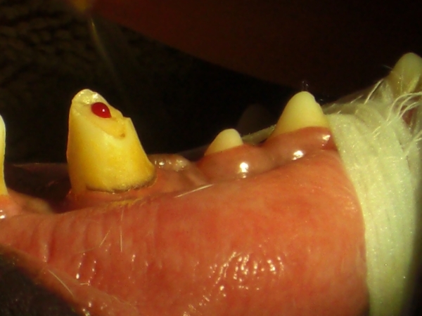
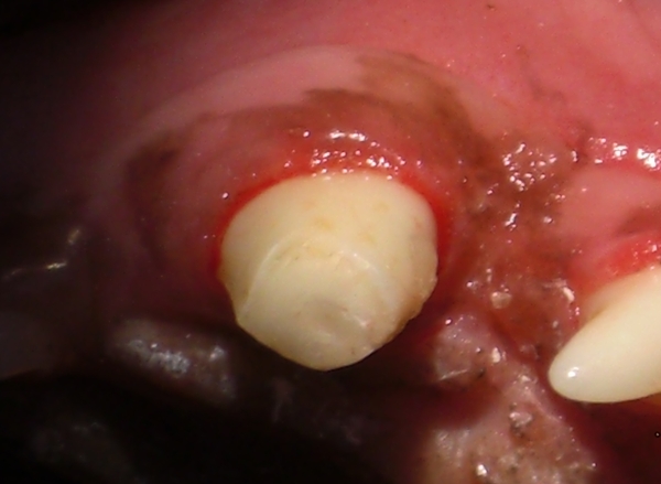
-

- Fractured tooth with bleeding pulp.
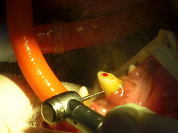
-

- Drilling of access hole for root canal.
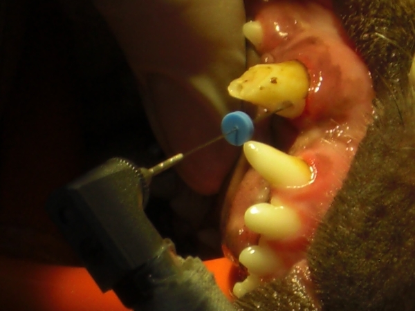
-

- The first cleaning of the root canal is done by hand with so-called hedstrom files.
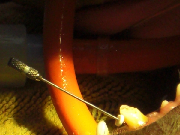
-

- Preparing the root canal with Lightspeed® rotary file system for filling.
-

- Damaged tooth after filling and sealing.
-

- X-ray of root canal to confirm the filling is completed properly.
